Cancer has been a driving force behind many of my research efforts, informing projects that span optical imaging, targeted contrast agent development, and in vivo sensing. These efforts have sought to address critical gaps across the cancer care continuum—from early detection to intraoperative guidance and longitudinal therapy monitoring—through the integration of optical technologies, spectroscopy, and nanoscale sensing platforms.
Early Diagnostics and Clinical Spectroscopy: Initial work focused on enabling early detection through a multi-modal spectroscopic platform for identifying epithelial precancers in the oral cavity and cervix. This system—later termed Fast Excitation-Emission Matrix (FastEEM)—integrated fluorescence and reflectance spectroscopy to acquire real-time, tissue-specific optical signatures (Technology in Cancer Research & Treatment, 2003). The approach supported optical biopsy without tissue removal, allowing rapid biochemical characterization in settings lacking histopathology. By advancing fast, label-free detection of early transformation, this work helped demonstrate the feasibility of noninvasive screening tools adaptable to primary care and low-resource settings. These methods laid important technical and conceptual foundations for downstream diagnostic platforms.
Targeted Contrast Agents for Cancer Imaging: To support molecularly specific detection of tumors, I led the development of multimodal protein-shell microspheres engineered with iron oxide nanoparticles, near-infrared fluorophores, and integrin-targeting ligands. These agents were optimized for in vitro and in vivo use with magnetomotive OCT, fluorescence imaging, and MRI (Cancer Research, 2010, 2011). Follow-up work enhanced tunability and signal amplification through nanoparticle seeding (IEEE JSTQE, 2018). These contrast agents demonstrated selective accumulation in tumors and lymph nodes across preclinical breast cancer models but were designed to be platform-agnostic across solid tumors. This work was supported by a U.S. Department of Defense Breast Cancer Research Program fellowship and helped advance a broader vision for real-time, multiplexed molecular imaging.
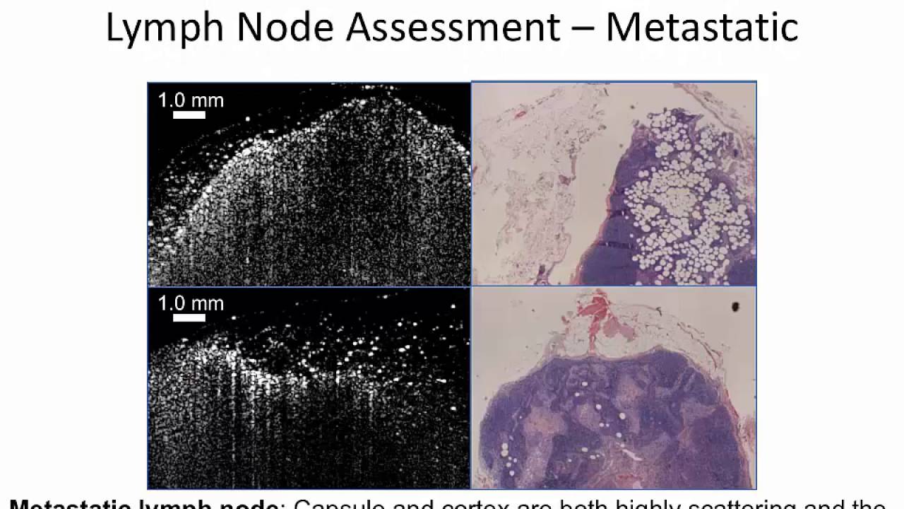
Intraoperative Imaging for Surgical Oncology: Recognizing the challenges of margin assessment in breast-conserving surgery, I developed portable OCT systems for real-time, high-resolution imaging during resection. These systems differentiated malignant and benign tissue using scattering signatures and microstructural features, and were deployed clinically in patients to evaluate tumor margins and lymph node architecture (Cancer Research, 2009; Journal of Biomedical Optics, 2010). This work also included the development of needle-based OCT probes for deeper tissue access and clinical integration. These efforts led to a 2015 U.S. patent (US 9,877,987) and contributed to a paradigm shift in surgical oncology by introducing label-free, real-time decision support at the point of care.
Spectroscopic Monitoring of Therapeutic Response: Optical spectroscopy techniques were also employed to evaluate cancer treatment responses at the molecular level. In studies of multiple myeloma and glioblastoma, Raman spectral changes were correlated with apoptotic and cytoskeletal remodeling following drug exposure (SPIE, 2016; Biomedical Optics Express, 2018). In parallel, SERS-active nanoparticles enabled high-resolution detection of subcellular changes in breast cancer cells (Nanoscale, 2015). These approaches provided sensitive, noninvasive readouts of treatment efficacy and contributed to a growing interest in using real-time optical signatures for therapy optimization. This work underscored the value of spectroscopy not only for diagnosis, but also for informing treatment strategies.
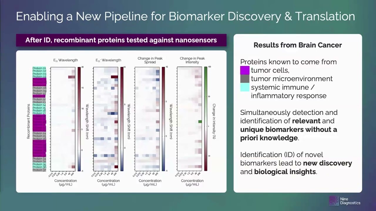
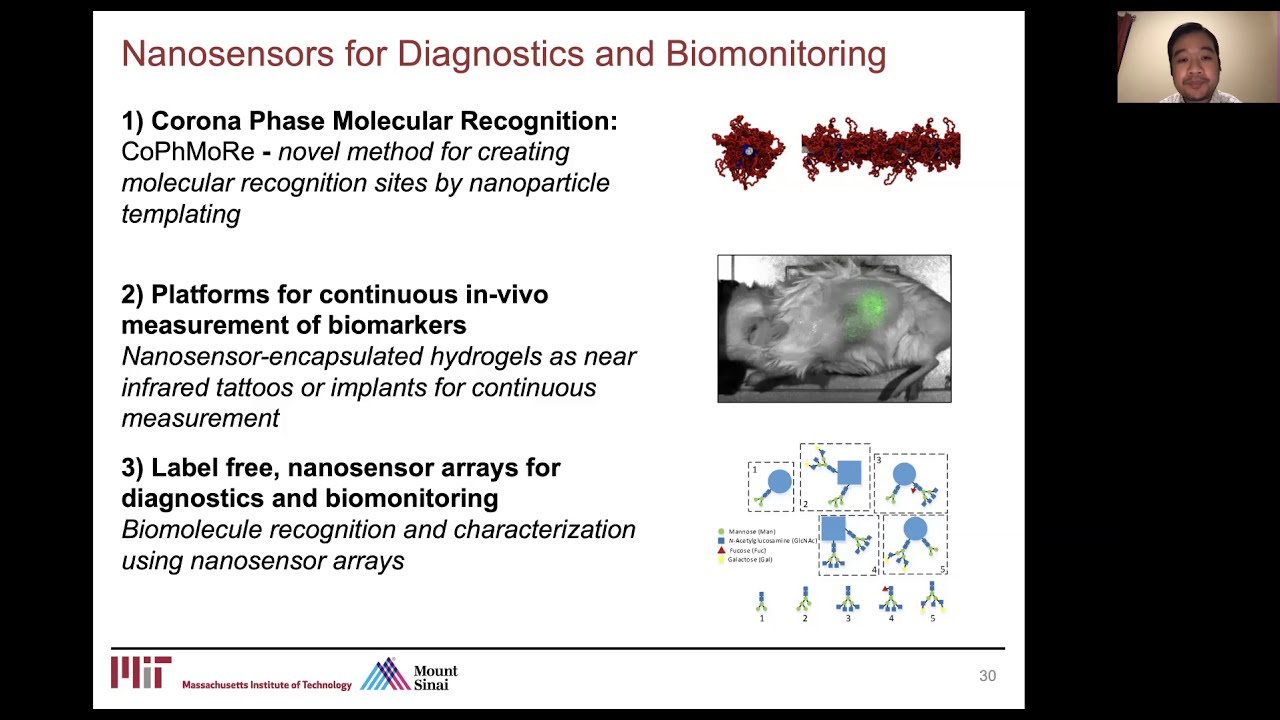
Nanosensors for In Vivo Disease and Treatment Monitoring: Most recently, I led the development of carbon nanotube–based nanosensors for real-time in vivo monitoring of oxidative stress and chemotherapeutic activity in tumor microenvironments. These sensors were applied in pancreatic ductal adenocarcinoma models to assess hydrogen peroxide levels as a marker of therapeutic response (Cancer Research, 2019). I also developed new sensors for monitoring drug pharmacokinetics and delivery, supported by wavelength-induced frequency filtering for autofluorescence suppression (Nature Nanotechnology, 2022) and a fiber-optic benchtop interface. This work—funded by an Arnold O. Beckman Postdoctoral Fellowship—resulted in a 2024 U.S. patent (US 11,969,021) and represents a step toward clinically integrated, molecularly targeted monitoring tools.
These translational innovation efforts link optical science, nanotechnology, and clinical insight to address cancer as a multifaceted, systems-level challenge. While much of this work has focused on breast, pancreatic, brain, cervical, oral, and gastrointestinal cancers, the tools and principles developed have broad applicability across both solid and hematologic malignancies. Across disciplines and teams, these contributions have supported a vision of precision oncology guided by molecular diagnostics, real-time imaging, and responsive therapeutic monitoring.


 of Cells and Tissues")


 as a diagnostic tool for the real-time intraoperative assessment of breast cancer surgical margins")


