Optical coherence tomography (OCT) has been a central focus of my research, enabling high-resolution, depth-resolved imaging for structural, biomechanical, and molecular characterization of tissue. My work spans the development of OCT systems, design of targeted contrast agents, and clinical translation for image-guided surgical decision-making—advancing OCT from a benchtop imaging technique to a point-of-care diagnostic tool.
System Development and Imaging Platform Engineering: To bring OCT into clinical workflows, I developed portable, real-time imaging systems with custom optics and integrated visualization tools. These platforms were optimized for high-speed data acquisition, ease of use, and intraoperative deployment, enabling clinicians to acquire high-resolution images during surgery or diagnostic procedures. By adapting OCT for handheld and cart-based operation, this work helped bridge the gap between optical imaging laboratories and the surgical suite—supporting real-time tissue assessment and procedural guidance in dynamic clinical environments. These early systems laid the technical foundation for OCT’s use in translational oncology and image-guided pathology.
Contrast Agents and Functional Imaging Capabilities: To extend OCT’s capabilities beyond structural imaging, I led efforts to develop targeted, multimodal contrast agents designed for magnetomotive OCT (MM-OCT). These microspheres integrated magnetic nanoparticles, near-infrared fluorophores, and integrin-targeting ligands to enable molecular specificity and biomechanical contrast (Cancer Research, 2010–2012). I also contributed to the use of magnetomotive optical coherence microscopy to quantify tissue viscoelastic properties, enabling biomechanical imaging at the microscale (SPIE, 2011). Building on this foundation, I led the development of nanoparticle-seeded microspheres with tunable magnetic response and signal enhancement, supporting advanced imaging and therapeutic monitoring applications (IEEE JSTQE, 2019). These functional extensions positioned OCT as a more informative modality, capable of resolving tissue architecture alongside molecular and mechanical biomarkers.
Clinical Translation and Intraoperative Application: To address critical needs in surgical oncology, I contributed to translating OCT into the operating room for real-time assessment of tumor margins and lymph node architecture. In breast-conserving surgery, OCT enabled differentiation of malignant from normal tissue using scattering intensity and microstructural features—allowing intraoperative margin assessment without destructive processing (Cancer Research, 2009; J Biomed Opt, 2010). These systems were deployed directly into surgical and pathology settings, demonstrating OCT’s potential to complement histopathology with rapid, high-resolution, and noninvasive tissue characterization.
My contributions have helped establish OCT as a versatile imaging platform that supports functional interrogation, surgical precision, and clinical decision-making at the point of care.

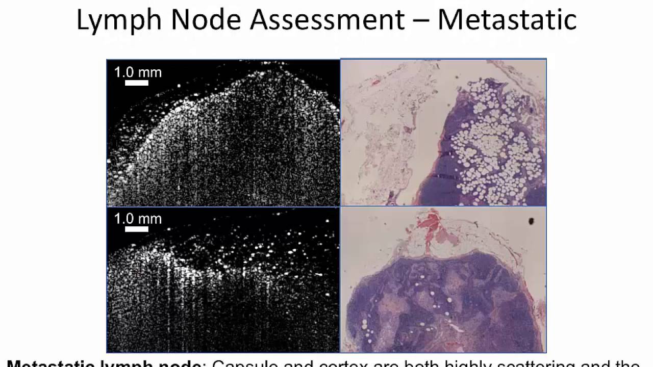


 as a diagnostic tool for the real-time intraoperative assessment of breast cancer surgical margins")


