Contrast agents are essential to modern imaging, enhancing resolution, specificity, and the ability to visualize biological and pathological processes noninvasively. My research has focused on the development of targeted, multimodal protein microspheres that enable integration across multiple imaging platforms, including magnetomotive optical coherence tomography (MM-OCT), fluorescence imaging, and magnetic resonance imaging (MRI), with a primary emphasis on cancer applications.
Targeted Microsphere Design and Molecular Specificity: The microsphere platform was built on a dual-phase structure: a hydrophobic oil core encapsulated within a hydrophilic protein shell. This structure allowed simultaneous loading of iron oxide nanoparticles and near-infrared fluorophores, creating a versatile vehicle for magnetic and optical contrast. Functionalization with RGD peptides enabled receptor-specific binding to αvβ3 integrins, which are overexpressed in tumor neovasculature. These agents were validated in vitro across a panel of breast cancer cell lines and in vivo in preclinical tumor models, showing selective accumulation at sites of neovascular growth. The goal was to detect molecular signatures of cancer progression before anatomical changes became apparent—an essential capability for early diagnosis and image-guided intervention.
Multimodal Imaging Integration and Functional Contrast: Once established as targeted probes, these microspheres were integrated into MM-OCT workflows to generate magnetically induced motion contrast in highly scattering tissues. This enabled visualization of tissue biomechanics and molecular composition within a single imaging session. Their compatibility with other modalities such as MRI and wide-field fluorescence imaging expanded the utility of the platform. Studies also demonstrated the use of magnetomotive optical coherence microscopy to capture intracellular responses at micrometer-scale resolution. Optimizations to the microsphere design—such as tunable size, improved magnetic response, and layered coatings—enhanced their performance across systems and became a central focus of later development efforts.
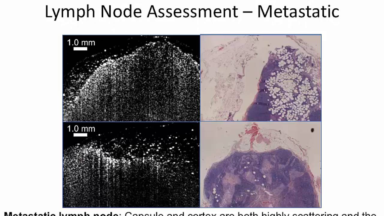
Translational Applications and Imaging Impact: Preclinical studies applied this platform to address real-world diagnostic needs, including tumor margin delineation, lymph node evaluation, and vascular targeting in breast cancer models. The ability to correlate optical, mechanical, and molecular signatures enabled more comprehensive assessments of tumor architecture. These agents demonstrated the potential to serve as a foundation for intraoperative imaging tools and to evolve into theranostic platforms capable of delivering therapy alongside imaging. By moving beyond passive enhancement, this work positioned contrast agents as active, tunable components in the advancement of image-guided medicine and precision diagnostics.
This body of work demonstrates how rationally engineered agents can redefine the role of contrast in next-generation imaging. Their modular design continues to support innovation at the interface of biology, materials science, and clinical translation.