Skip to content
Freddy T. Nguyen, MD, PhD
Research Fellow @ Massachusetts Institute of Technology
CEO, Co-Founder @ Nine Diagnostics
Google Scholar
PubMed
ResearchGate
ORCID
Dimensions.AI
Google Scholar
PubMed
ResearchGate
ORCID
Dimensions.AI
X-twitter
Linkedin
Instagram
Facebook
Threads
Home
About
News
Media
Press
Mentions
Publications
Peer-Reviewed
Reviews
Patents
White Papers
Innovation
Patents
Nine Diagnostics
Prevented Health
MIT Healthcare Innovation
MIT Hacking Racism Challenge
MIT COVID-19 Challenge
MIT Hacking Medicine
Project Prana Foundation
wePool.AI
Physician Scientist
American Physician Scientists Association
People
Photonics
Fluorescence Spectroscopy
Optical Coherence Tomography
Raman Spectroscopy
Photonics
Fluorescence Spectroscopy
Optical Coherence Tomography
Raman Spectroscopy
Nanotechnology
Carbon Nanotubes
Contrast Agents
Nanotechnology
Carbon Nanotubes
Contrast Agents
Medicine
Cancer
Transfusion Medicine
Medicine
Cancer
Transfusion Medicine
Physician-scientist developing biophotonics and nano technologies for functional precision medicine to provide the right treatment to the right patient at the right time.
Massachusetts Institute of Technology
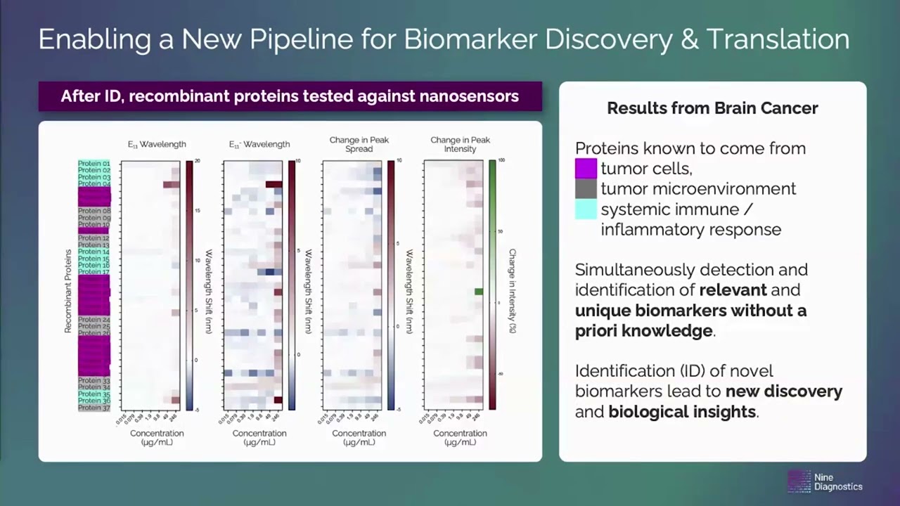
2025 MIT Health Science Forum: Treatment Effectiveness, New Biology Using AI – Enabled Nanosensor Technology (MIT Industrial Liaison Program – Startup Exchange)
May 8, 2025
Nine Diagnostics Joins American Cancer Society’s BrightEdge Entrepreneurs Program
February 12, 2025
Nine Diagnostics Selected for Merck Digital Sciences Studio Cohort 3
November 14, 2024
Freddy Nguyen, MD/PhD and Nine Diagnostics Win Novo Nordisk Golden Ticket at Pitch Event
October 8, 2024
Nine Diagnostics Wins Novo Nordisk Golden Ticket for LabCentral Residency
September 19, 2024
Fluorescence-based detection of protein aggregation and fiber optic-based benchtop instrument
August 6, 2024
MIT Fellow Freddy Nguyen, MD/PhD, on Precision Medicine and Fostering Physician-Scientist Trainees
February 3, 2024
MIT Healthcare Innovation
January 1, 2024
Laser Fluence Dependent Modulation of Single Walled Carbon Nanotube Photoluminescence
August 28, 2023
Molecular Recognition and In Vivo Detection of Temozolomide and 5-Aminoimidazole-4-carboxamide for Glioblastoma Using Near-Infrared Fluorescent Carbon Nanotube Sensors
December 16, 2022
Emerging technologies in cancer detection
June 17, 2022
Nature Nanotechnology: A wavelength-induced frequency filtering method for fluorescent nanosensors in vivo
May 30, 2022
Previous
Page
1
Page
2
Page
3
Page
4
Page
5
Page
6
Page
7
Page
8
Page
9
Next