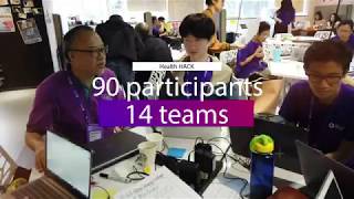
MIT Hong Kong Innovation Node – HealthHACK 2019
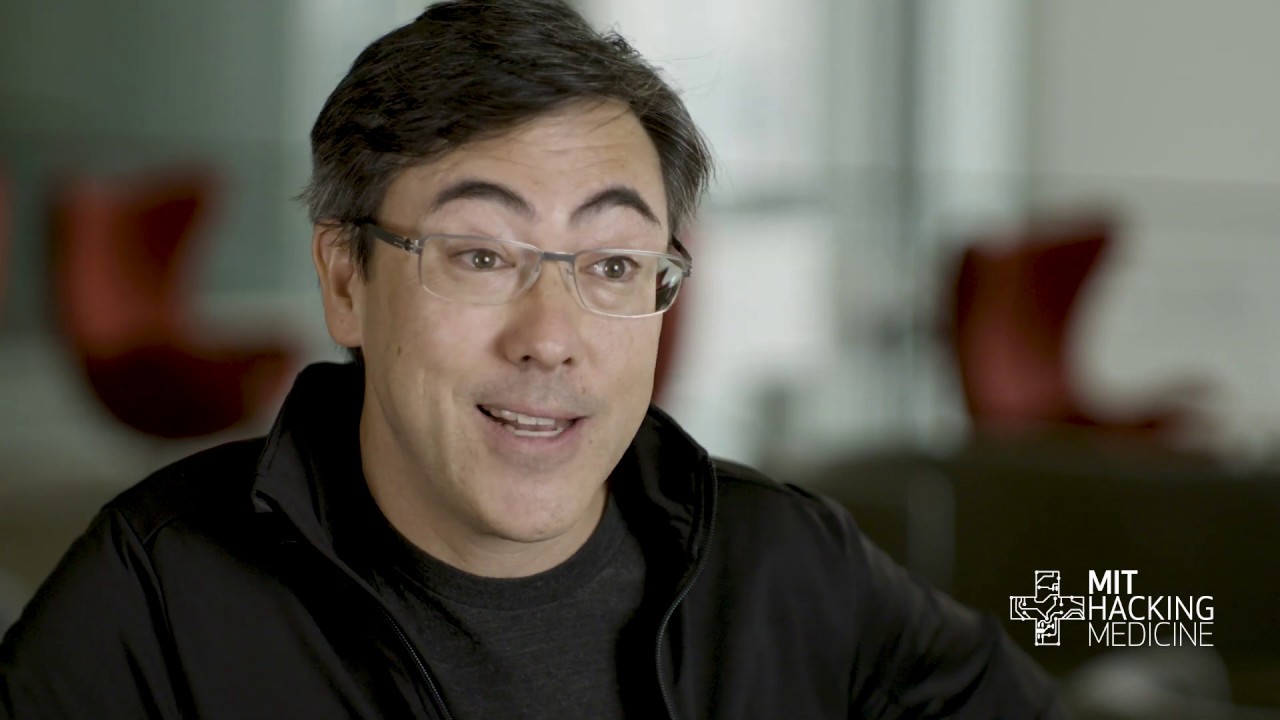
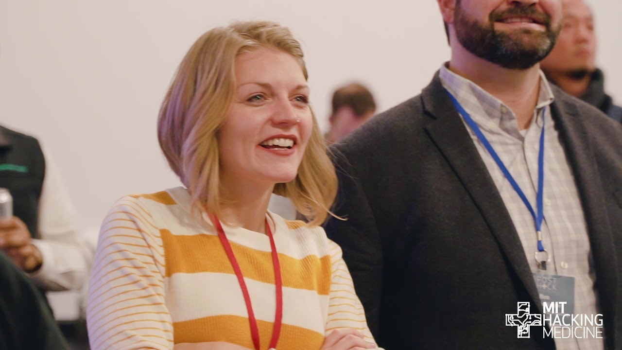
MIT Hacking Medicine – Boston Grand Hack 2019
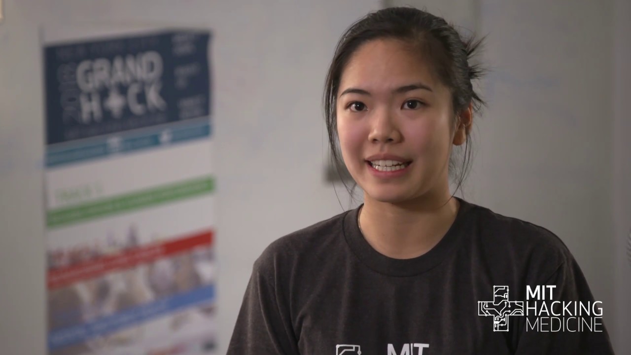
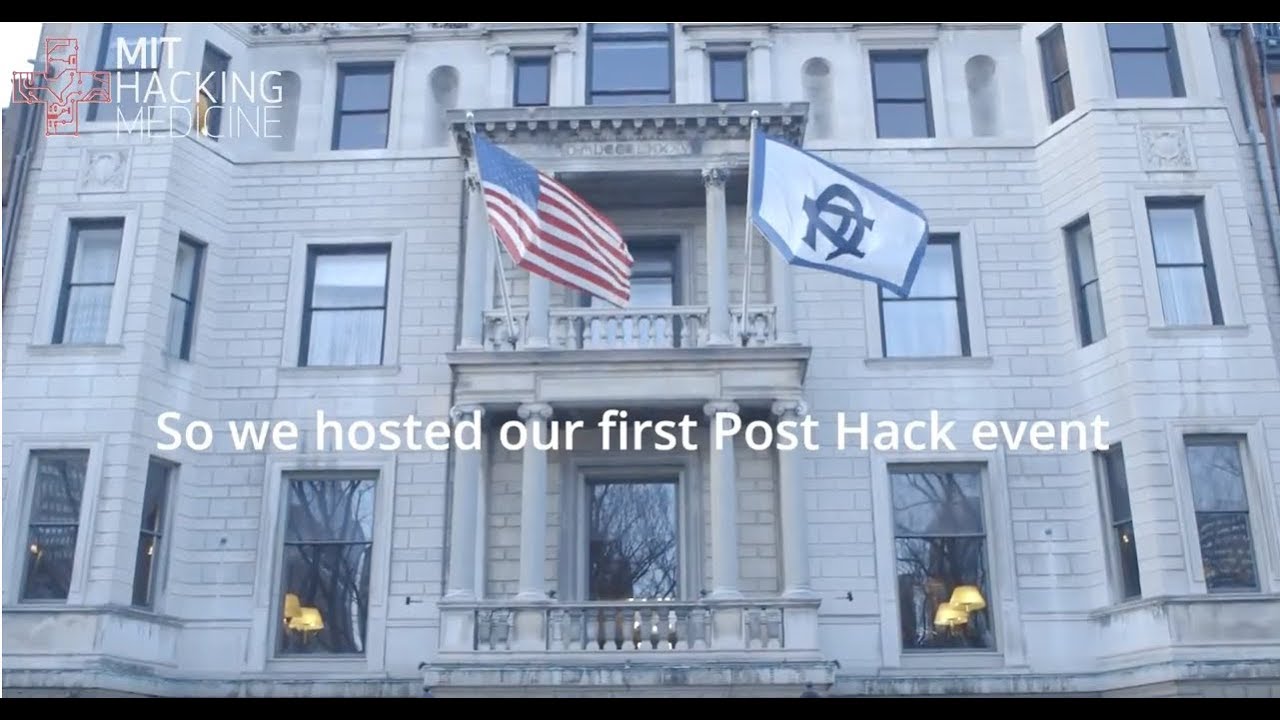
MIT Hacking Medicine – Post-Hack 2019
Implanted Nanosensors in Marine Organisms for Physiological Biologging: Design, Feasibility, and Species Variability

In recent decades, biologists have sought to tag animals with various sensors to study aspects of their behavior otherwise inaccessible from controlled laboratory experiments. Despite this, chemical information, both environmental and physiological, remains challenging to collect despite its tremendous potential to elucidate a wide range of animal behaviors. In this work, we explore the design, feasibility, and data collection constraints of implantable, near-infrared fluorescent nanosensors based on DNA-wrapped single-wall carbon nanotubes (SWNT) embedded within a biocompatible poly(ethylene glycol) diacrylate (PEGDA) hydrogel. These sensors are enabled by Corona Phase Molecular Recognition (CoPhMoRe) to provide selective chemical detection for marine organism biologging. Riboflavin, a key nutrient in oxidative phosphorylation, is utilized as a model analyte in in vitro and ex vivo tissue measurements. Nine species of bony fish, sharks, eels, and turtles were utilized on site at Oceanogràfic in Valencia, Spain to investigate sensor design parameters, including implantation depth, sensor imaging and detection limits, fluence, and stability, as well as acute and long-term biocompatibility. Hydrogels were implanted subcutaneously and imaged using a customized, field-portable Raspberry Pi camera system. Hydrogels could be detected up to depths of 7 mm in the skin and muscle tissue of deceased teleost fish ( Sparus aurata and Stenotomus chrysops) and a deceased catshark ( Galeus melastomus). The effects of tissue heterogeneity on hydrogel delivery and fluorescence visibility were explored, with darker tissues masking hydrogel fluorescence. Hydrogels were implanted into a living eastern river cooter ( Pseudemys concinna), a European eel ( Anguilla anguilla), and a second species of catshark ( Scyliorhinus stellaris). The animals displayed no observable changes in movement and feeding patterns. Imaging by high-resolution ultrasound indicated no changes in tissue structure in the eel and catshark. In the turtle, some tissue reaction was detected upon dissection and histopathology. Analysis of movement patterns in sarasa comet goldfish ( Carassius auratus) indicated that the hydrogel implants did not affect swimming patterns. Taken together, these results indicate that this implantable form factor is a promising technique for biologging using aquatic vertebrates with further development. Future work will tune the sensor detection range to the physiological range of riboflavin, develop strategies to normalize sensor signal to account for the optical heterogeneity of animal tissues, and design a flexible, wearable device incorporating optoelectronic components that will enable sensor measurements in moving animals. This work advances the application of nanosensors to organisms beyond the commonly used rodent and zebrafish models and is an important step toward the physiological biologging of aquatic organisms.
MIT Hacking Medicine – New York City Grand Hack 2018
In vivo detection of drug-induced apoptosis in tumors using Raman spectroscopy

We describe a label-free approach based on Raman spectroscopy, to study drug-induced apoptosis in vivo. Spectral-shifts at wavenumbers associated with DNA, proteins, lipids, and collagen have been identified on breast and melanoma tumor tissues. These findings may enable a new analytical method for rapid readout of drug-therapy with miniaturized probes.
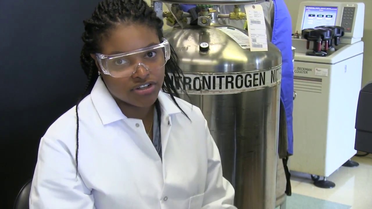
Beckman Foundation: 2018 Beckman Symposium Video Highlight Reel
Children’s Hospital of Philadelphia – Mitochondrial Research Affinity Group Seminar
Characterization of magnetic nanoparticle-seeded microspheres for magnetomotive and multimodal imaging

Magnetic iron-oxide nanoparticles have been developed as contrast agents in magnetic resonance imaging (MRI) and as therapeutic agents in magnetic hyperthermia. They have also recently been demonstrated as contrast and elastography agents in magnetomotive optical coherence tomography and elastography (MM-OCT and MM-OCE, respectively). Protein-shell microspheres containing suspensions of these magnetic nanoparticles in lipid cores, and with functionalized outer shells for specific targeting, have also been demonstrated as efficient contrast agents for imaging modalities such as MM-OCT and MRI, and can be easily modified for other modalities such as ultrasound, fluorescence, and luminescence imaging. In addition to multimodal contrast-enhanced imaging, these microspheres could serve as drug carriers for targeted delivery under image guidance. Although the preparation and surface modifications of protein microspheres containing iron oxide nanoparticles has been previously described and feasibility studies conducted, many questions regarding their production and properties remain. Since the use of multifunctional microspheres could have high clinical relevance, here we report a detailed characterization of their properties and behavior in different environments to highlight their versatility. The work presented here is an effort for the development and optimization of nanoparticle-based microspheres as multi-modal contrast agents that can bridge imaging modalities on different size scales.
MIT Hacking Medicine – Post-Hack 2018
2017 Class of Arnold O. Beckman Postdoctoral Fellows

The Arnold and Mabel Beckman Foundation announced today its 2017 class of Arnold O. Beckman Postdoctoral Fellows, individuals who underscore the Foundation’s mission of supporting basic research in the chemistry and life sciences. They were selected after a three-part review led by a panel of scientific experts.
The Foundation will award more than $2.6 million in funding for 20 exceptional research fellows from 13 universities
MIT News: Developing rapid cancer nano sensors

Chemicals like nitric oxide and hydrogen peroxide can promote cancer growth. MPC-CMSE Summer Scholar Kaila Holloway is working in the lab of Michael S. Strano, Carbon P. Dubbs Professor in Chemical Engineering at MIT, to develop tiny chemical sensors to detect their concentrations near tumors in the body.
